Do not routinely offer imaging for uncomplicated low back pain
BMJ 2021; 372 doi: https://doi.org/10.1136/bmj.n291 (Published 12 February 2021) Cite this as: BMJ 2021;372:n291Linked Editorial
Sustainable practice: what can I do?

- Amanda M Hall, assistant professor1,
- Kris Aubrey-Bassler, director1,
- Bradley Thorne, patient advisor1,
- Chris G Maher, director2
- 1Primary Healthcare Research Unit, Faculty of Medicine, Memorial University of Newfoundland, St John’s, Newfoundland A1B 3V6, Canada
- 2Institute for Musculoskeletal Health, University of Sydney and Sydney Local Health District, Sydney, Australia
- Correspondence to: A M Hall amanda.Hall{at}med.mun.ca
What you need to know
Less than 5-10% of all low back pain is due to a specific underlying spinal pathology
The remaining 90-95% has no indication of a serious cause and should be managed with conservative treatments such as advice and reassurance, exercise, physical therapy, chiropractic care, cognitive-behavioural therapy, or pain management
Diagnostic triage based on clinical history and examination can help distinguish between non-specific or more serious low back pain
Imaging may do more harm than good when serious conditions are not suspected and is likely to prolong recovery in patients with non-specific low back pain
Patients’ primary concerns of whether their pain is caused by something serious and what they should do to aid recovery can be addressed by sound education and reassurance, without the need for imaging
The clinical problem
The past two decades have seen a paradigm shift in the way we use imaging when managing low back pain (LBP). Imaging was once a routine part of the diagnostic workup for most cases of LBP. Evidence now indicates that imaging is useful only in the small subgroup of patients for whom there is suspicion of red flag conditions. These conditions include cancer, infection, inflammatory disease, fracture, and severe neurological deficits—which together account for only 5-10% of LBP presentations in primary care.1 For the remaining 90-95% of LBP cases (called non-specific or uncomplicated LBP), imaging will not guide management and can cause more harm than benefit. International guidelines2 and “Choosing Wisely” campaigns now encourage a diagnostic triage approach to identify those patients who require imaging (box 1). Given these advances in knowledge, imaging rates for LBP should be decreasing, but recent systematic reviews show the opposite, reporting that imaging has increased over the past 20 years3 and that at least a third of all images are unnessary.4
“Choosing Wisely” statements from different countries related to avoiding the use of routine imaging for low back pain
“Don’t routinely image patients with low back pain regardless of the duration of symptoms unless: (a) there are clinical reasons to suspect serious underlying pathology (i.e., red flags), or (b) imaging is necessary for the planning and/or execution of a particular evidenced-based therapeutic intervention on a specific spinal condition.”—Canadian Spine Society, Choosing Wisely Canada
“Do not undertake imaging for low back pain in patients without indications of a serious underlying condition.”—Australian Rheumatology Association, Choosing Wisely Australia
“Do not routinely offer imaging in a non-specialist setting for people with low back pain with or without sciatica. Consider imaging in specialist settings of care (for example, a musculoskeletal interface clinic or hospital) for people with low back pain with or without sciatica only if the result is likely to change management.”—Royal College of Physicians, Choosing Wisely UK
The evidence for change
Evidence from randomised controlled trials has established that imaging does not improve clinical outcomes, and several observational studies have linked liberal imaging with greater work absence and unnecessary use of health services.5 Unnecessary LBP imaging has the potential for harm. Perhaps the most obvious of these harms is exposure to radiation from x rays and computed tomography. Harm is also possible if a clinician acts on incidental findings from any imaging method, including magnetic resonance imaging (MRI), or if these findings are not appropriately explained to patients. For example, disc abnormalities such as bulging disc or degenerative disc disease are commonly seen on images but may not be the source of pain, as they are also seen on images in up to 97% of asymptomatic patients (table 1).6 Incidental findings may lead to further investigations, specialist referral, and more intensive treatment such as surgery, which limits access to those services for the patients who are in genuine need of such care.7 Incidental findings may also provoke worry or concern in patients from a labelling effect (possibly delaying recovery) if they fail to understand that these anomalies are common and benign.89
Age-specific prevalence estimates of degenerative spine imaging findings in asymptomatic people6
Imaging and follow-up based on incidental findings affects the health system in terms of direct costs (such as cost of equipment, staff time to conduct the imaging, and time to interpret the image findings) and downstream costs (subsequent tests, referrals, and interventions, many of which are expensive and are of limited benefit for most patients with back pain). The downstream costs of imaging also affect patients and society due to indirect costs relating to out of pocket expenses for unnecessary interventions, as well as expenses related to lost work or household productivity.810 Two systematic reviews of six randomised controlled trials (1804 participants) both concluded that patients who received imaging without a clear clinical indication did not have improvements to pain, function, or quality of life compared with those who did not have imaging.1112 Additionally, observational studies have found that those who received imaging when not indicated had greater use of health services such as injections and surgery and greater work absence than their counterparts who did not receive imaging.5131415 For example, a 2020 study of 405 965 US primary care patients found that those who had early MRI were more likely to undergo back surgery (1.48% v 0.12%) and take prescription opioids (35.1% v 28.6%), yet had higher pain score at 1-year follow-up (3.99 v 3.87) than those who did not get early MRI.16
Barriers to change
Lack of awareness of current back pain guidelines and knowledge about how to apply them in practice are likely contributors to the over-reliance on imaging in primary care. This is often exacerbated if institutional policies and practices around imaging are not aligned to the latest evidence. Recent systematic reviews suggest that the clinical environment itself may encourage physicians to over-order imaging due to (a) accommodating patient requests, (b) believing that imaging will reassure the patient, and (c) not having time to explain and justify a non-imaging approach.17 Reported less consistently in the literature, additional factors include worry about missing a specific pathology, not being aware of conservative interventions besides medication, and a lack of access to conservative management such as exercise therapy, physical therapy, chiropractic care, cognitive behavioural therapy, and pain management programmes.1718
Patient perspectives
Clinicians often report that they order imaging when patients specifically request it. Systematic reviews of surveys and interviews suggest that about half of patients expect imaging from their health provider because they believe it can help rule out a sinister cause for the pain.19 Some patients also believe that imaging can identify the cause better than the clinician’s physical exam and facilitate a more tailored approach to treatment.19 There is still considerable uncertainty as to what alternatives to imaging would convey the diagnosis of non-specific LBP satisfactorily from the patient’s perspective. Future research should focus on this area to develop strategies for explaining LBP to patients.
How should practice change?
Practice changes in the following key areas are required to support appropriate imaging use:
Diagnostic triage and management—Evidence suggests that, by conducting a more thorough diagnostic triage,20 clinicians can discern which patients fall into the category of non-specific LBP. Papers by Bardin et al20 and Traegar et al21 present a useful visual aid (fig 1) for conducting diagnostic triage in patients with LBP. Bardin et al’s visual aid also details evidence based treatments for patients assessed to have radicular syndrome or non-specific LBP. We have supplemented this with a decision support tool (fig 2) to help clinicians identify the small subgroup of patients with suspected red flag conditions who do need imaging. As detailed in figure 1, advise all patients with non-specific LBP or radicular syndromes, regardless of pain duration, to remain active and reassure them of a good prognosis.

Diagnostic triage for low back pain. (Adapted from Traegar et al21)

Decision aid based on the latest evidence for triaging LBP patients. (Imaging for all other conditions is probably not useful and may be harmful.) (Adapted from Bardin et al20)
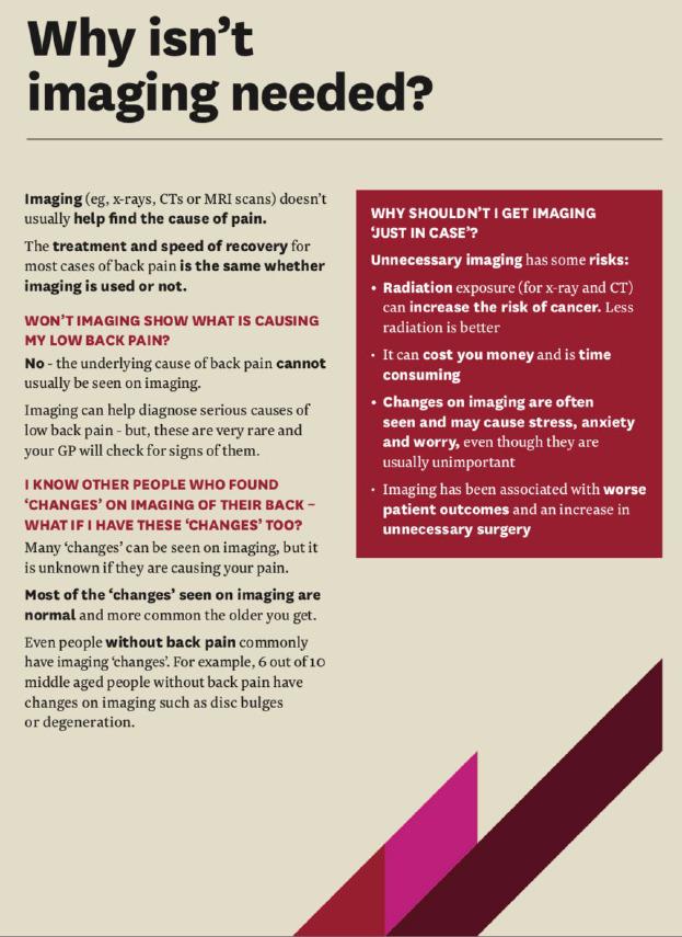
Patient education—Offer patients with non-specific LBP reassurance that imaging is not required and provide advice on self management as first-line care. Box 2 summarises key points to cover when providing reassurance. Jenkins et al’s education booklet Understanding My Low Back Pain guides clinicians and patients through points to cover within a consultation22 and can be given to patients as an educational and treatment advice tool (see fig 3 for an example).
What patients need to know
Most cases of LBP are simple strains and sprains of the back that, while painful and unpleasant, improve rapidly just like a sprained ankle
Having an image (x ray or CT or MRI scan) of your back does not usually help to find the cause of the back pain or guide treatment
The treatment for most cases of back pain is the same whether imaging is used or not; and we have seen that those who have unnecessary imaging often have a delayed recovery
Unnecessary imaging has some risks, including radiation exposure and delay in appropriate treatment, and has been associated with worse patient outcomes and an increase in unnecessary surgery
You may know other people who have had an x ray, CT, or MRI of their back that showed “changes”; but most of the changes seen on imaging are normal and are more common the older you get, just like grey hair and wrinkles. As these changes also occur in people without back pain, their relevance is unclear

Screenshot from patient education booklet Understanding My Low Back Pain provided as a data supplement by Jenkins et al22 (available at: https://www.ncbi.nlm.nih.gov/pmc/articles/PMC6154885/bin/12913_2018_3526_MOESM3_ESM.pdf)
Communication style—Findings from a recent qualitative study suggests four key behaviours from clinicians that can help patients feel reassured.23
Show that you “know the whole story” by summarising the patient’s medical history and conducting a thorough assessment
Demonstrate empathy and communicate that you are qualified and experienced, helping the patient to feel that they are “seeing the right person”
Reduce the use of generic statements such as “nothing to worry about” and recognise the patient’s distress
Explain the likely cause(s) of LBP and provide a clear self management plan to help the patient understand and manage their LBP.22
Monitoring—Regularly monitor image ordering practices and LBP outcomes relative to peers. This will reassure clinicians that the changes to practice that they implement are effective and do not result in patient harm.
Search methods
We searched Medline and the Cochrane Library to identify published systematic reviews on (i) the use of LBP imaging, (ii) the concordance of imaging with guidelines, (iii) physician reported barriers to guideline adherence, (iv) patient expectations around LBP imaging, and (v) interventions that have been used to change LBP imaging practice. Additionally, we consulted experts in LBP and primary care on relevant published evidence and the most up to date LBP guidelines internationally.
Education into practice
How will your referrals for imaging in people with low back pain change as a result of reading this article?
Audit the number of spinal x rays and MRIs requested in your practice in the past six months. How many of those imaging requests are in line with the recommendations given in the diagnostic triage tool in fig 1?
How might you go about providing reassurance that imaging is not required?
How patients were involved in the creation of this article
Bradley Thorne is a patient advisor for the Primary Healthcare Research Unit Musculoskeletal Programme at Memorial University, Newfoundland. In this role, he has consulted on several projects related to improving the management of LBP. He has a special interest in back pain due to lived experiences with low back pain over many years. He reviewed a range of publicly available patient facing materials, as well as the published literature that informed the “Patient perspectives” section in addition to reviewing and commenting on subsequent versions of the manuscript.
Footnotes
This article is part of a series of Education articles based on recommendations from international Choosing Wisely campaigns. The BMJ thanks Wendy Levinson and Karen Born at Choosing Wisely for valuable advice and supporting the selection of topics. Choosing Wisely had no input into the peer review process or editorial decision.
We thank Helen Richmond (postdoctoral fellow) and Andrea Pike (senior research assistant) at the Faculty of Medicine, Memorial University for their assistance with searching the literature and reviewing the manuscript. We thank Wendy Levinson and Karen Born for suggesting the review topic and reviewing the manuscript.
Contributors: AH conducted the initial literature review (which was reviewed by KA and CM) and prepared the first draft of the article. BT reviewed the result from the literature search relating to the patients perspective. BT also reviewed the “Patients perspectives” section and reviewed literature specific to patients and public knowledge of and attitudes toward back pain imaging. KA and CM reviewed and revised the manuscript. AH is the guarantor for the paper.
Competing interests: We have read and understood the BMJ Group policy on declaration of interests and have no relevant interests to declare.
Provenance and peer review: Commissioned, based on an idea from the author; externally peer reviewed.
This is an Open Access article distributed in accordance with the Creative Commons Attribution Non Commercial (CC BY-NC 4.0) license, which permits others to distribute, remix, adapt, build upon this work non-commercially, and license their derivative works on different terms, provided the original work is properly cited and the use is non-commercial. See: http://creativecommons.org/licenses/by-nc/4.0/.